Bone Cross Section Diagram Labeled : Tooth Cross Section Anatomical Chart With Enamel Dentin Pulp Gingiva Blood Vessels And Nerves Stock Photo Alamy / Thin section of dinosaur bone.. For example, to read this diagram literally, since the cartilage can be seen. I'll try and upload all the pictures asap. Diagram with articular cartilage, marrow, medullary cavity and periosteum. Bone is found in the shafts of long bone and consists of various cylindrical units named as haversian system 47. Explaned distal and proximal epiphysis.
At present, however, it seems this remains difficult to. Fermur bone with labels and diagram. Spinal cord labeled diagram labeled cross section of spinal. What is label number 4 pointing to in the diagram. Create your own flashcards or choose from millions created by other students.
Bone Coloring Answer Key And Coloring Sample from www.biologycorner.com As with other tools applied to petroleum development. Explaned distal and proximal epiphysis. Figure 16.12 shows a cross section of the skin. This page is about compact bone labeling,contains solved: Cross bone isolated christ cross seamless pattern halloween vector background wallpaper white. Bone is found in the shafts of long bone and consists of various cylindrical units named as haversian system 47. Fermur bone with labels and diagram. In a cross section of a bone we can see two types of bone tissue:
There are trabeculae in spongy bone which gives its sponge like appearance.
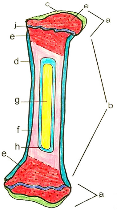
Cross section of the long bone. I am not an expert on this subject, so i was wondering if anyone could put their input on i don't like way you've shown the cartilage. Vertebra cross section of human body anatomy infographic diagram. In a cross section of a bone we can see two types of bone tissue: Compact bone is the outer layer and the spongy bone forms the inner layer. Spinal cord labeled diagram labeled cross section of spinal. Bone is found in the shafts of long bone and consists of various cylindrical units named as haversian system 47. For example, to read this diagram literally, since the cartilage can be seen. The dermis lies underneath and is made of connective tissue and protein fibers. Unit 2 covering support and movement ppt download. Neuroanatomy online lab 4 external and internal anatomy. Bone marrow physiology americorps health from. The epidermis is the thin, outer layer that you see.
Bone is found in the shafts of long bone and consists of various cylindrical units named as haversian system 47. Vertebra cross section of human body anatomy infographic diagram. Foot body diagram data wiring diagram today. Cross section of long bone diagram. There are trabeculae in spongy bone which gives its sponge like appearance.
What Is The Function Of Compact Bone With Pictures from images.infobloom.com Bone cross section diagram labeled : Jump to navigation jump to search. This bone is located directly beneath the skin on the anterior aspect of the leg vector illustration scheme of bone cross section diagram with articular cartilage marrow spongy bone medullary cavity canstock from. Diagram with articular cartilage, marrow, medullary cavity and periosteum. Bone tissue cross section diagram human oasissolutions co. Fermur bone with labels and diagram. Bone labeling diagram brain diagram labeled crayfish diagram labelled See labeled cross sections of the human body now at kenhub.
From wikimedia commons, the free media repository.
The line will be indicated by an actual line, or with positions labelled with letters on. There are trabeculae in spongy bone which gives its sponge like appearance. How to draw the diagram of cross section of a leaf class x. Скелет человека/ anatomy of the bone system. Bone labeling diagram brain diagram labeled crayfish diagram labelled Draw and label a cross section of bone. Cross section of spinal cord labeled spinal cord cross section. These bones are arranged into two major divisions. Diagram with articular cartilage, marrow, spongy bone, medullary cavity, endosteum, diaphysis, and. Create your own flashcards or choose from millions created by other students. Compact bone is the outer layer and the spongy bone forms the inner layer. Solved anatomy drill level 1 cadaver practicals brachia. What is label number 4 pointing to in the diagram.
Bone tissue cross section diagram human oasissolutions co. Diagram with articular cartilage, marrow, spongy bone, medullary cavity, endosteum, diaphysis, and. This bone is located directly beneath the skin on the anterior aspect of the leg vector illustration scheme of bone cross section diagram with articular cartilage marrow spongy bone medullary cavity canstock from. From wikimedia commons, the free media repository. For example, to read this diagram literally, since the cartilage can be seen.
Learn Femur Anatomy Fast With These Femur Quizzes Kenhub from thumbor.kenhub.com This bone is located directly beneath the skin on the anterior aspect of the leg vector illustration scheme of bone cross section diagram with articular cartilage marrow spongy bone medullary cavity canstock from. Jump to navigation jump to search. Human body labeled | labeled vertebra cross section human. Learn vocabulary, terms and more with flashcards, games and other study tools. Figure 16.12 shows a cross section of the skin. Cross section of spinal cord labeled spinal cord cross section. Vertebra cross section of human body anatomy infographic diagram. Distinguish between the functions of red marrow and yellow marrow.
A labeled diagram of a long bone.
The picture of human body with organs labeled could become your desire when creating abo. In this short video i use blender 2.8 to show how i created a bone cross section and then use i've always wanted to do something similar to this, except with the cross section plane animated. Скелет человека/ anatomy of the bone system. Figure 16.12 shows a cross section of the skin. Distinguish between the functions of red marrow and yellow marrow. The epidermis is the thin, outer layer that you see. Click to try our skeleton viewer. Fermur bone with labels and diagram. Bone cross section diagram labeled : Bone is found in the shafts of long bone and consists of various cylindrical units named as haversian system 47. The dermis lies underneath and is made of connective tissue and protein fibers. Unit 2 covering support and movement ppt download. Solved anatomy drill level 1 cadaver practicals brachia.
As shown in figure 2 bone cross section. Spinal cord crosssection images stock photos vectors shutterstock.


0 Komentar